Cellular and Molecular Anatomy of Cilia

Eukaryotic cilia are microtubule-based organelles that project from the cell surface to drive motility, signaling, and sensing. Nearly all vertebrate cells are ciliated, bearing a primary cilium, and, in specialized epithelia, are multiciliated, containing tens to hundreds of motile cilia for coordinated flow. The same cilia that cover vertebrate cells are structurally remarkably similar to those of unicellular organisms spanning eukaryotic clades, suggesting the last eukaryotic common ancestor bore a cilium. The conservation across evolution offers an opportunity to use structural cell biology to understand the similarity and nuances between species and develop general principles that exist across evolution.
The Cellular and Molecular Anatomy of Cilia lab will focus on understanding the cellular structural biology and dynamics of cilia. We will use a combination of in situ structural biology techniques in several model systems to address questions regarding how cells build and maintain cilia, the evolution of ciliary dynamics, ciliary adaptation, intracellular ciliary heterogeneity, and how all of these themes tie into our understanding of ciliopathies.
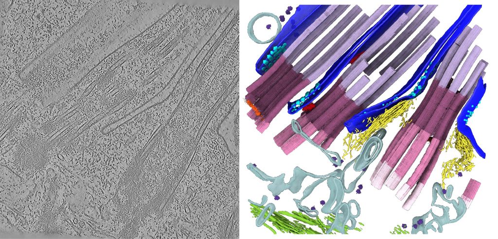
In situ structural cell biology
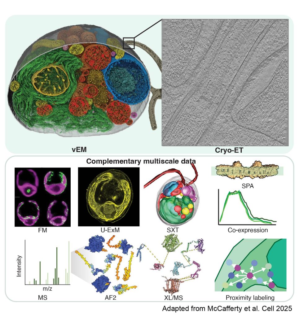
Integration of multimodal data
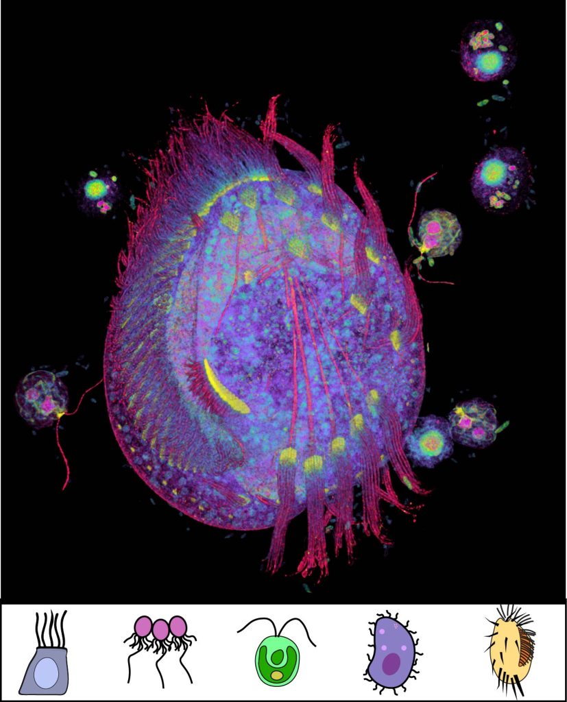
Evolution of ciliary dynamics & heterogeneity
Publications
- Molecular architecture of the ciliary base in mammalian multiciliated cells, 2026
- Loss-of-function variants in SAXO6, encoding a microtubule inner protein of photoreceptor cilia, cause a late-onset retinal dystrophy, 2026
- Stoic: Fast and accurate protein stoichiometry prediction, 2026
- Toward community-driven visual proteomics with large-scale cryo-electron tomography of Chlamydomonas reinhardtii, 2026
- The CAGE complex: a hollow, megadalton, protein assembly in prokaryotic and eukaryotic microbes, 2025
- The luminal ring protein C2CD3 acts as a radial in-to-out organizer of the distal centriole and appendages, 2025
- An amino acid-resolution interactome for motile cilia identifies the structure and function of ciliopathy protein complexes, 2025
- Integrating cellular electron microscopy with multimodal data to explore biology across space and time, 2024
- Integrated modeling of the Nexin-dynein regulatory complex reveals its regulatory mechanism, 2024
- Does AlphaFold2 model proteins’ intracellular conformations? An experimental test using cross-linking mass spectrometry of endogenous ciliary proteins, 2023
- Native doublet microtubules from Tetrahymena thermophila reveal the importance of outer junction proteins, 2023
- Integrative modeling reveals the molecular architecture of the intraflagellar transport A (IFT-A) complex, 2022
- The protein organization of a red blood cell, 2022
- Improving integrative 3D modeling into low-to medium-resolution electron microscopy structures with evolutionary couplings, 2021
- Simplified geometric representations of protein structures identify complementary interaction interfaces, 2021
- Structural Biology in the Multi-Omics Era, 2020